MEG
Magnetoencephalography (MEG)
Neuromag Vector View
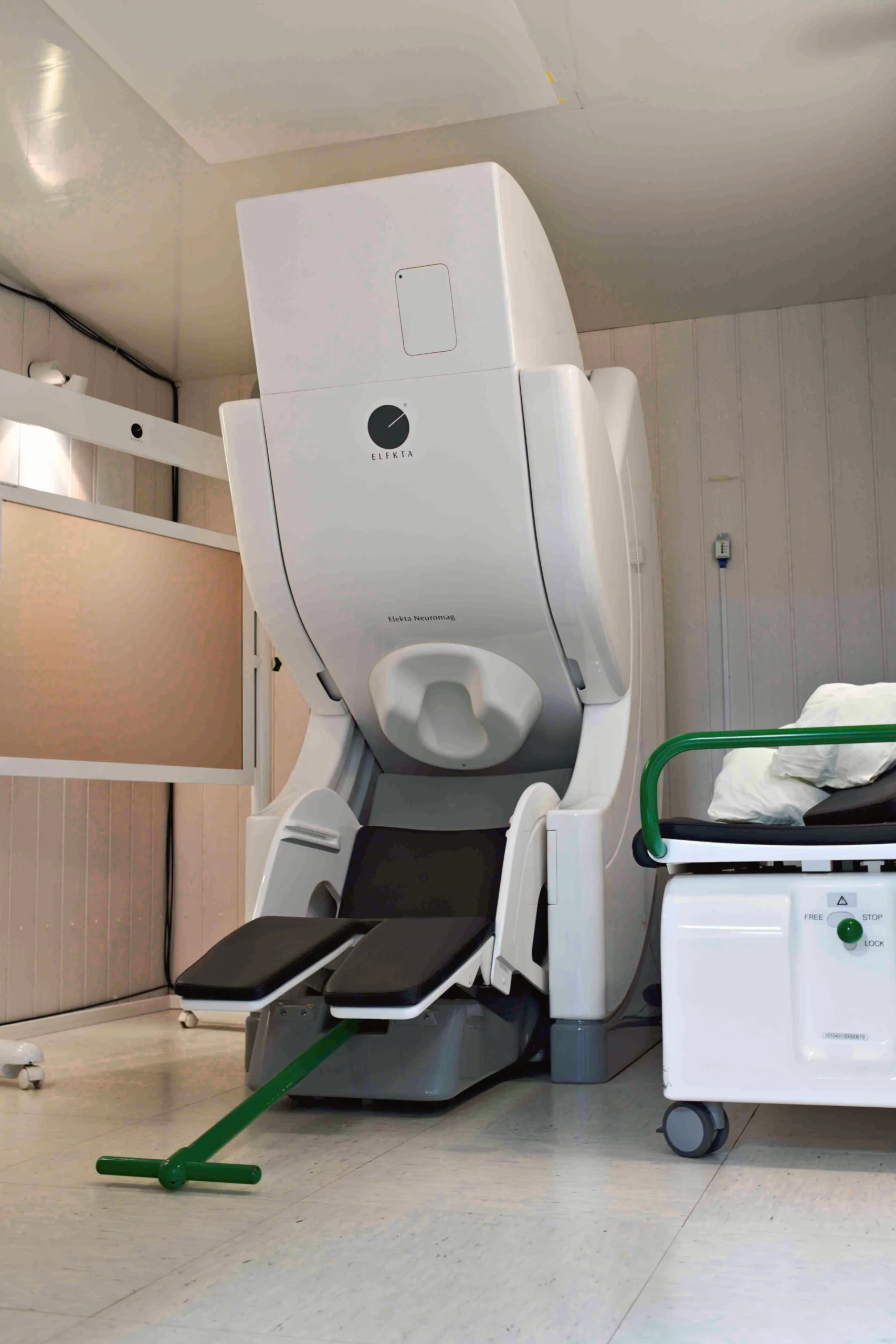
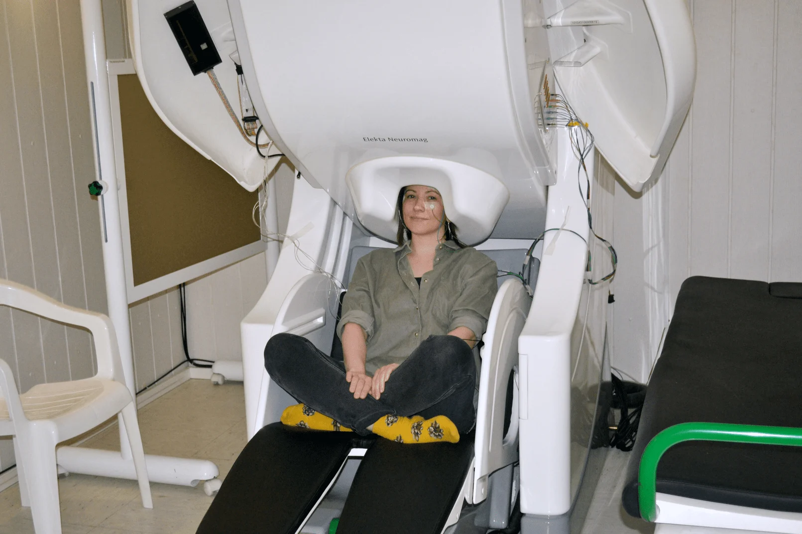
The core system at the MEG Center is the 306-channel magnetoencephalograph ‘Neuromag Vector View’ (Elekta Oy, Finland).
Manuals:
Magnetically Shielded Room
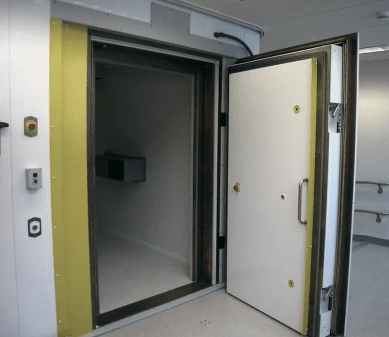
The MEG system is located inside a two-layer magnetically shielded room “Ak3B” (Vacuum-schmelze GmbH, Germany), which suppresses external magnetic fields and makes possible registration of the tiny electromagnetic fields of the brain.
Electroencephalography (EEG)
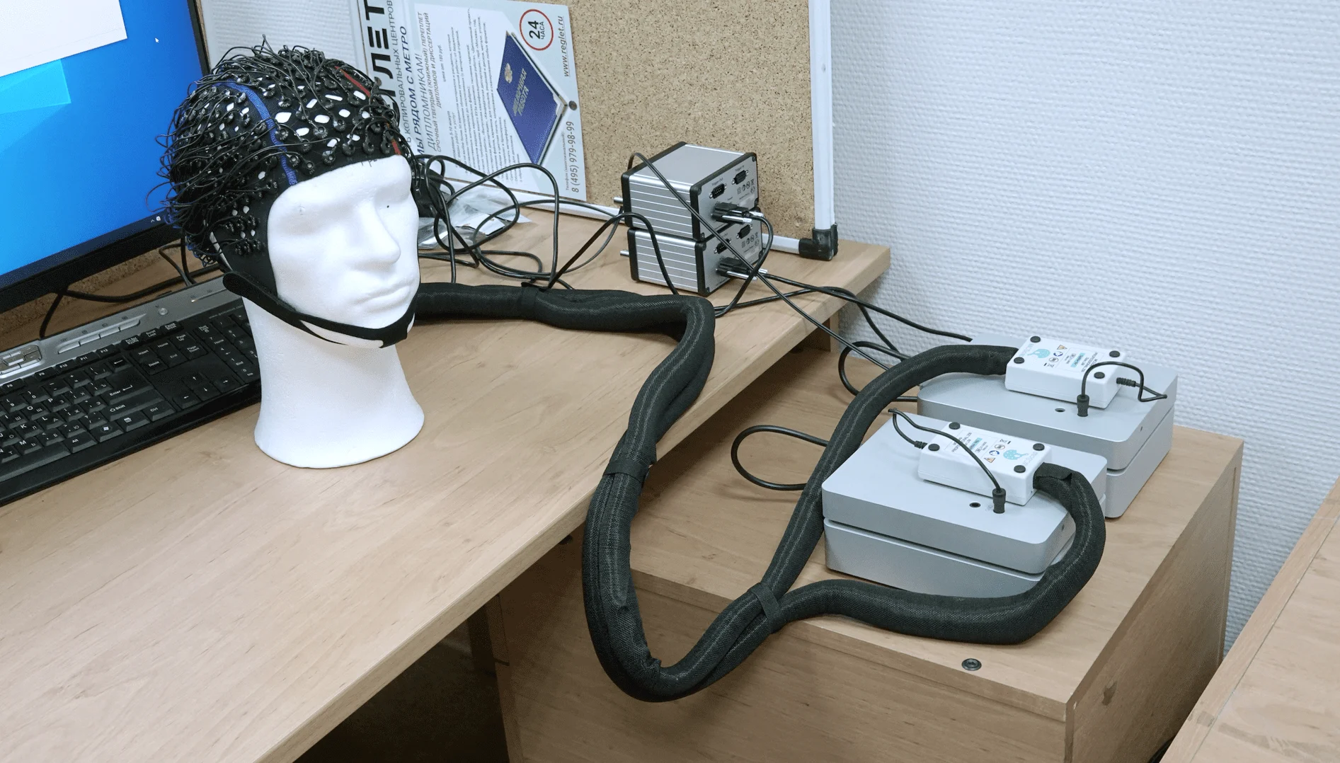
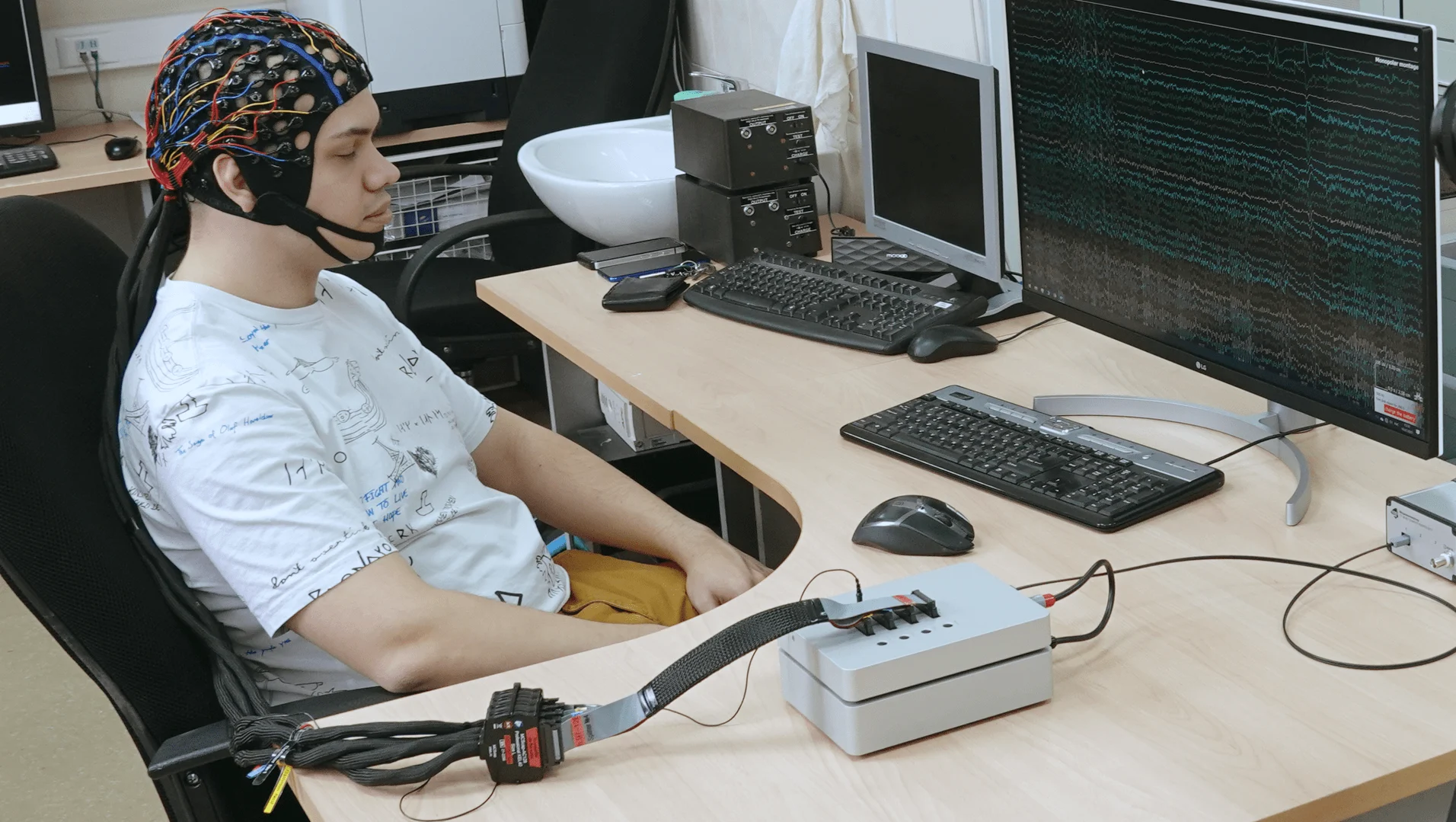
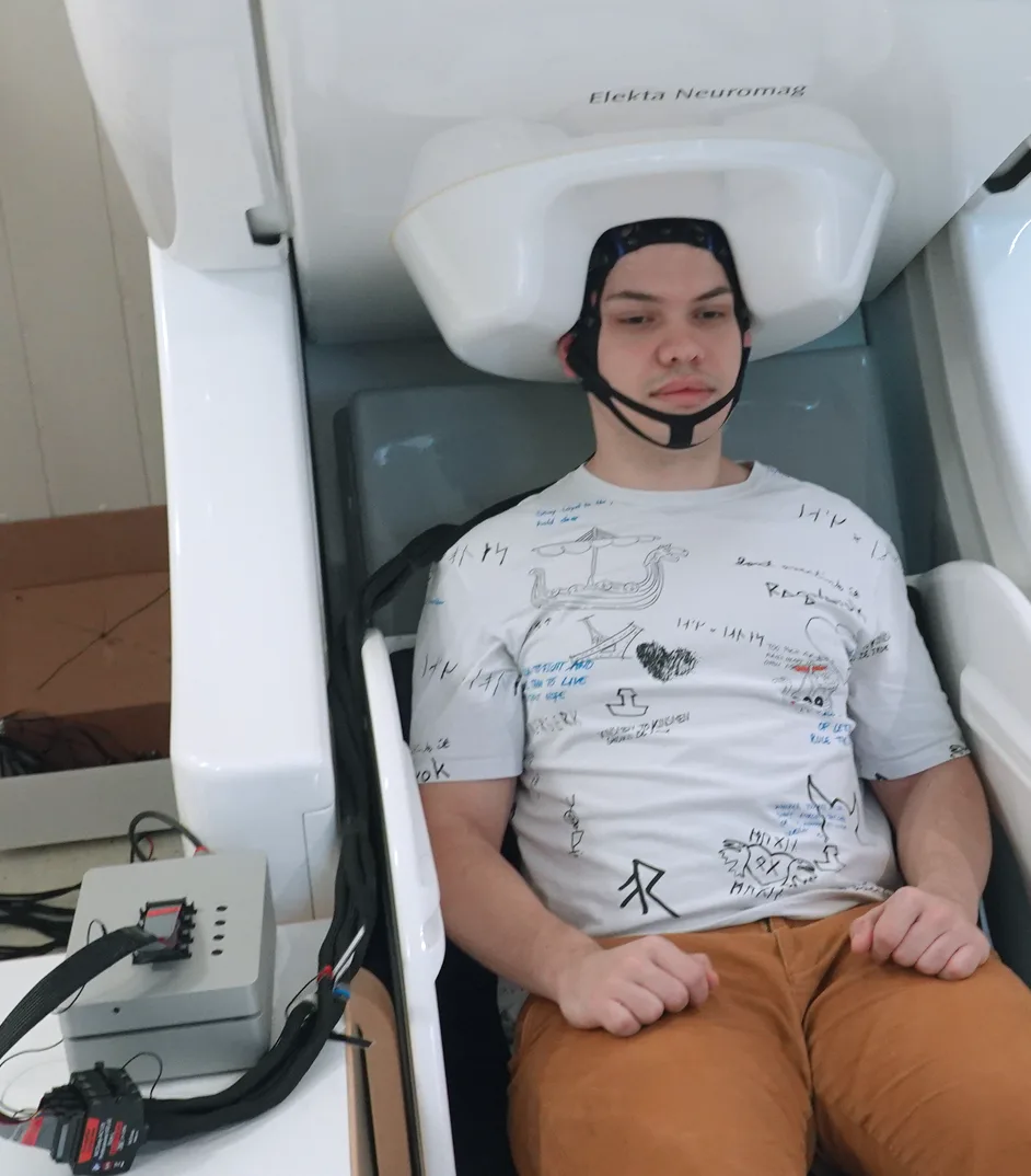
We have several systems for EEG registration, which allow you to choose the optimal solution depending on the experimental task.
In combination with other equipment, such as magnetoencephalograph, magnometers with optical pumping, high-precision MEG-compatible eyetracker EyeLink 1000 Plus, etc., these electroencephalographs allow you to create a variety of equipment complexes.
For EEG studies not related to synchronous registration of EEG and MEG we use a separate grounded shielded room, specially designed by Russian specialists.
EEG system “NVX-136” with an extension kit
Purchased in 2021 MEG-compatible EEG system “NVX-136” (Medical Computer Systems (MCS)) allows to register up to 128 channels (up to 256 channels with the expansion kit). EEG registration is possible both separately and synchronously with the MEG.
An autonomous 32-channel system “ActiCHamp” for recording EEG, ECG and EMG
The autonomous 32-channel “ActiCHamp” system for recording EEG, ECG and EMG (Brain Products GmbH, Germany) is equipped with active electrodes, which allow to significantly reduce the noise level. The “ActiCHamp” system allows registration of the EEG separately and synchronously with the MEG.
A 40-channel system “Neocortex-Pro”
Electroencephalograph and hardware-software complex “Neocortex-Pro” (Neurobotics, Russia) allow to register and analyze EEG.
Optically Pumped Magnetometers
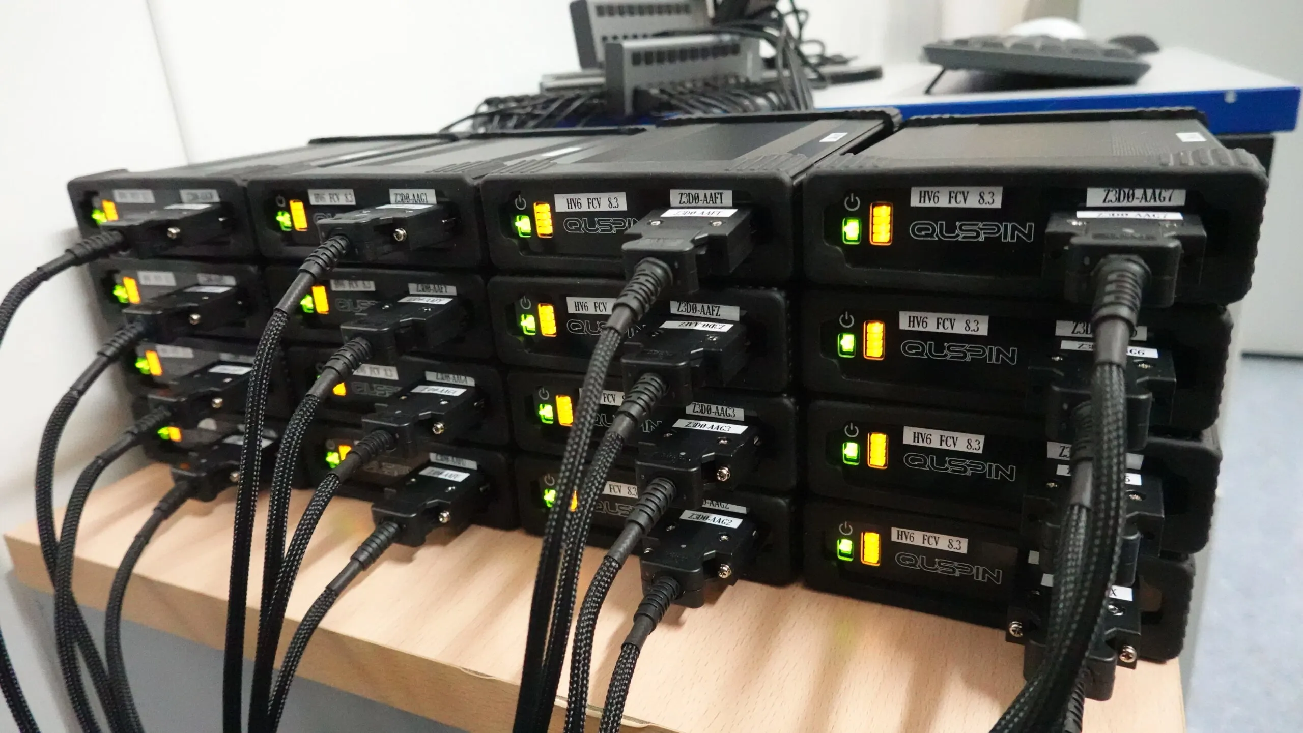
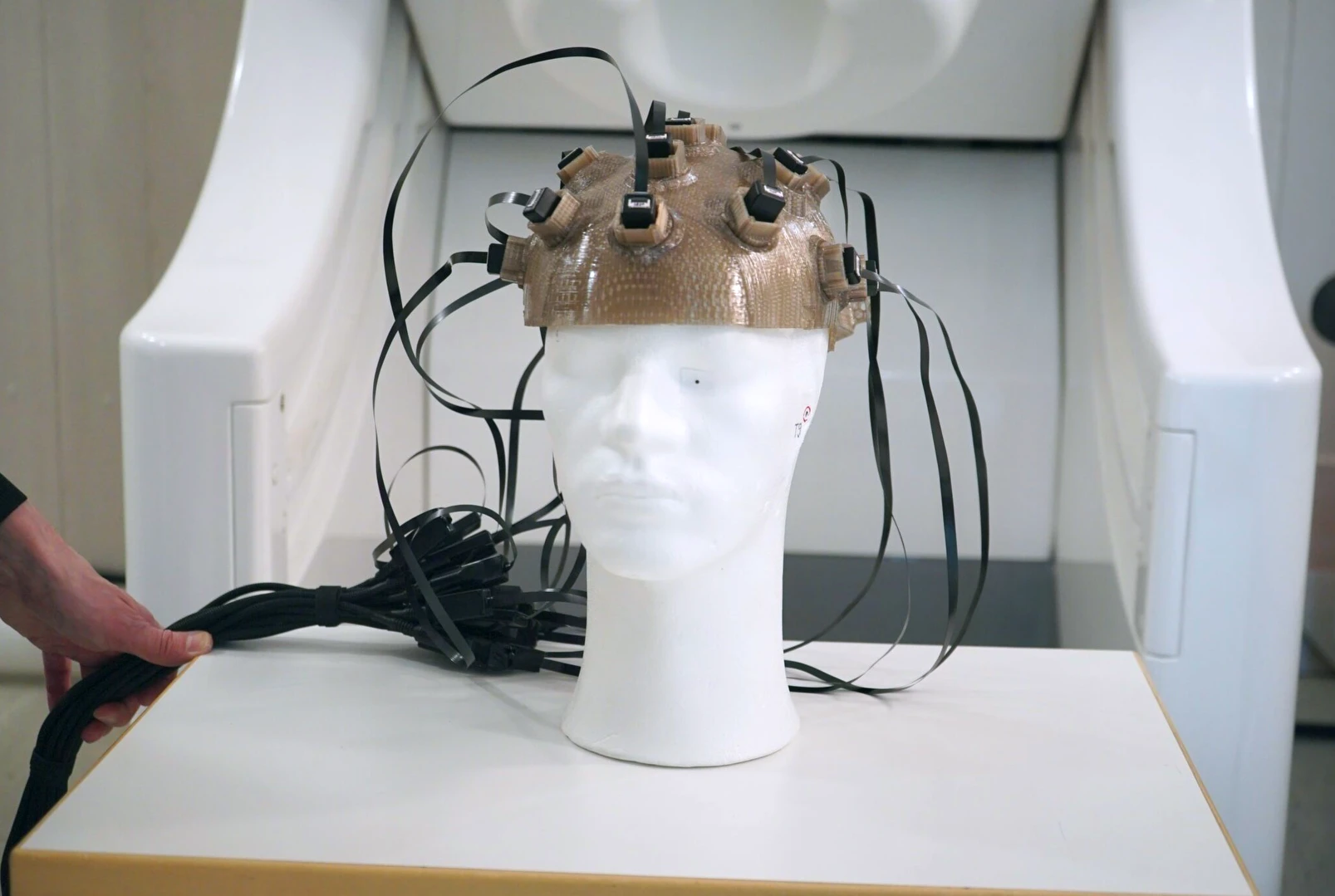
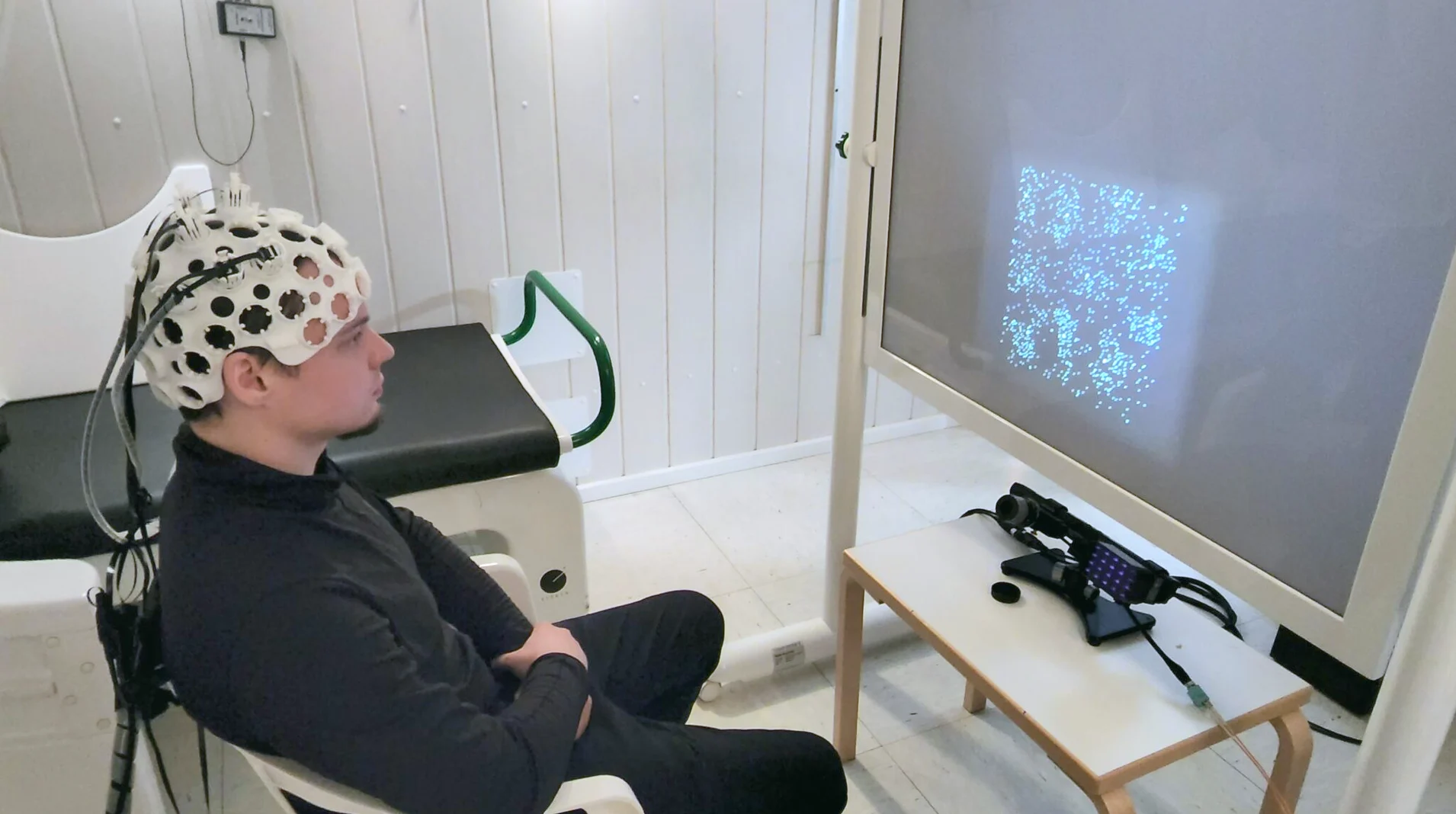
16 compact optically pumped magnetometers (OPM) QZFM Gen-3.0 (Quspin, USA, 2021 г) were purchased for the MEG-center in 2021.
The high–temperature portable quantum OPM is a unique technology for non-invasive study of the human brain that has appeared in recent years and is rapidly gaining popularity in the world. OPMs don’t require helium cooling and can be mounted directly on the participant’s head. This makes it possible to achieve a particularly high accuracy in the localization of the active areas of the brain and creates prospects for the portable magnetoencephalography devices.
For research with OPMs, we use a magnetically shielded room Ak3B, a 3d-printer for making individual helmets and other accessories. To create models of individual helmets and other accessories, an Artec Eva 3d scanner with Artec Studio 16 software was purchased in 2022.
Eye Tracker «EyeLink 1000 plus»
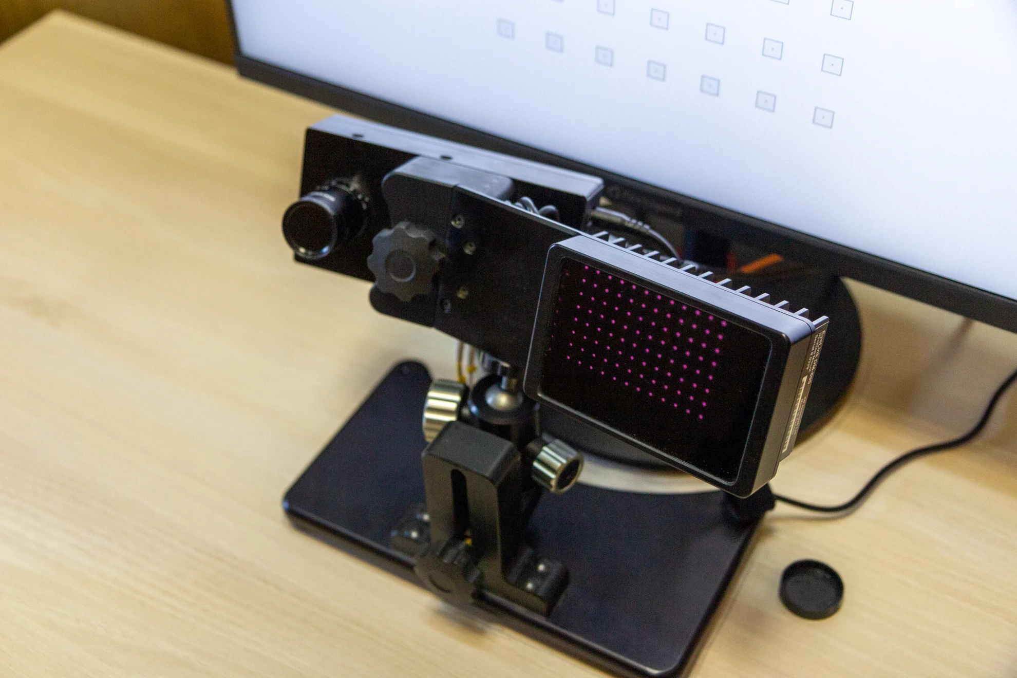
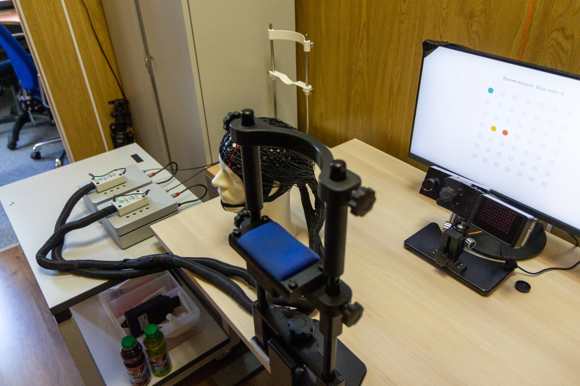
To monitor direction of the gaze, and to record saccades, fixations, and changes in pupil size, we use EyeLink-1000 plus eyetracker (SR Research Ltd., Canada). The system makes it possible to conduct oculomotor behavior studies separatly or simultaneously with MEG recording.
Visual Stimulation
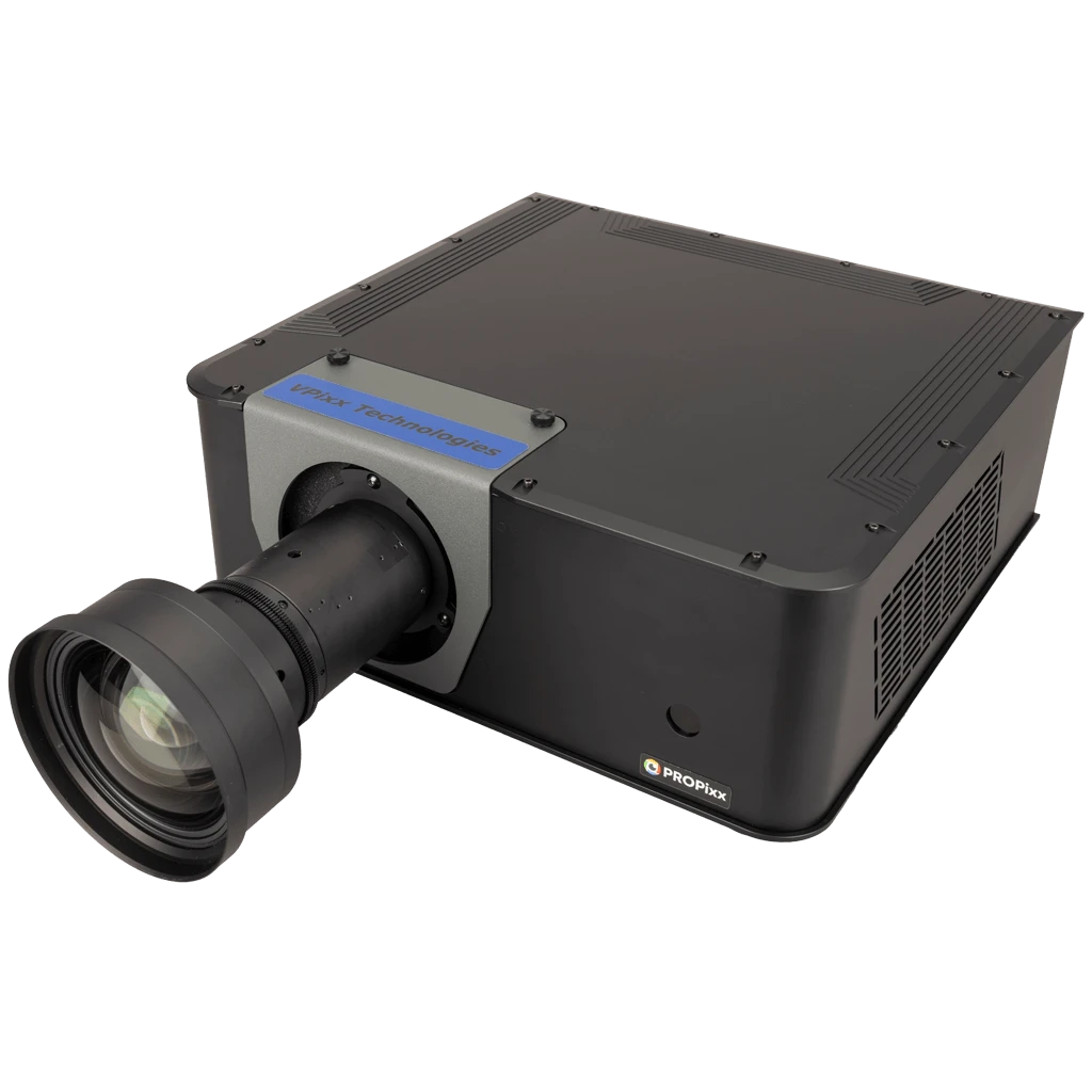
PROPIxx Lite
The PROPIxx Lite projector (VPixx Technologies Inc., Canada) features a high frame rate (up to 1440 Hz in Grayscale mode and up to 500 Hz in RGB mode), which allows you to:
- avoid imposing a standard frame rate on brain activity;
- create a more correct illusion of movement on presentation of moving stimuli;
- carry out frequency tagging of the parts of the visual stimulus, which is necessary for reliable control of the visual attention focus movement (including latent visual attention, independent of eye movements).
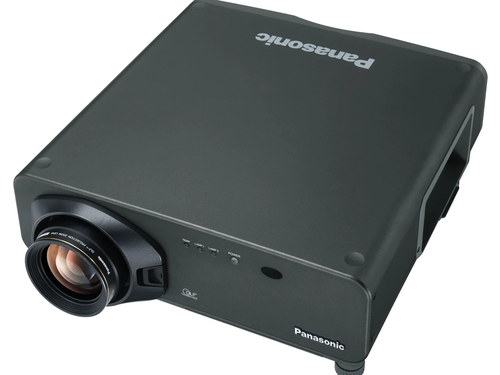
Panasonic PT-D7700E-K
For studies that don’t require high frequency visual stimuli we use the projector «Panasonic PT-D7700E-K».
Audio Stimulation
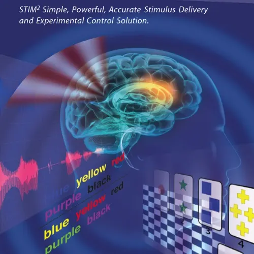
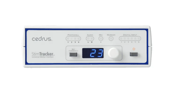
To apply auditory stimuli we use MEG-compatible audio system «NeuroScan STIM2» (Compumedics, USA).
STIM2 is fully integrated with the Cedrus StimTracker system, which utilizes photodetectors and auditory threshold detection to ensure maximum accuracy in stimulus timing.
STIM2 includes 14 modules, among them the universal programmable experiment control module Gentask, an audio file editor, and an image converter. The other modules cover motor, perceptual, cognitive, mnemonic, and attentional functions, including neuropsychological tests such as Stroop, Card Sorting, and others. Gentask enables the development and presentation of virtually any visual, auditory, or combined tasks, as well as the recording of behavioral responses. It is used in studies of evoked potentials (P300, N400, MMN, CNV) and visual memory tests. Built-in editors simplify working with audio files and graphic formats (BMP, JPG, PNG, TIFF), ensuring convenient preparation of stimulus materials.
Electro Stimulation
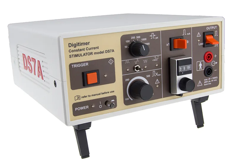
For electric stimulation of the median nerve, we use two electric stimulators «Digitimer DS7A» (USA).
The Digitimer DS7A high-voltage stimulator provides isolated constant current stimulation at 0-100 mA from a voltage source up to 400 V.
The DS7A features precise control elements for pulse duration (50 μs to 2 ms) and amplitude (0-100 mA), and includes external trigger options via either a foot/hand switch or a digital (TTL-compatible) trigger. This means that in addition to single stimulation pulses, the DS7A can be used for repetitive stimulation applications with frequencies up to 1 kHz (depending on current and pulse duration settings).
Tactile Stimulation
For tactile stimulation, we use fiber-optic devices developed at the MEG-center.
Capnograph
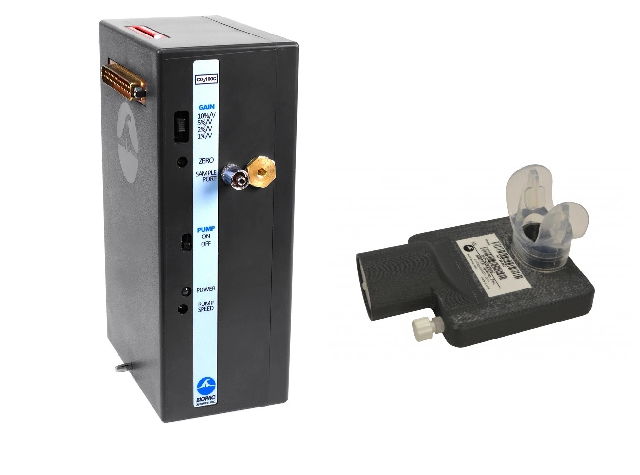
Hyperventilation is a convenient model for studying excitatory-inhibitory balance parameters, which is important in research on ASD and other neuropsychiatric disorders. During hyperventilation, a decrease in the CO₂-content on exhalation corresponds to an increase in the CO₂-concentration in the blood (alkalosis). Since the effect of alkalosis is non-linear, the CO₂-content on exhalation should be controlled during the experiment. For these purposes we have the capnograph (BIOPAC Systems Inc., USA).
3D Printer
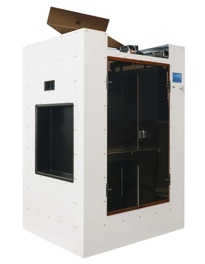
For studies with high-temperature optically pumped magnetometers, it is necessary to manufacture individual helmets and other accessories.
The MEG-center is equipped with Maestro Grand 2 FDM 3D printer (Show Design LLC, Russia), which provides work with a wide range of materials. Two extruders allow to print models of complex configuration.
Helium Recycling System
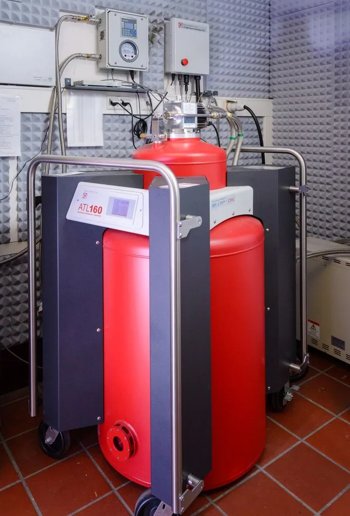
The functioning of the superconductive MEG sensors (‘squeeds’) requires cooling them to a temperature close to the absolute zero. Therefore, the sensors are placed in liquid helium. The helium recycling system “ATL160” of Quantum Design company (USA) liquefies the evaporating gas and provides for its re-use, which significantly reduces expenses for maintaining the system in a working state.
Registration of Motor Responses
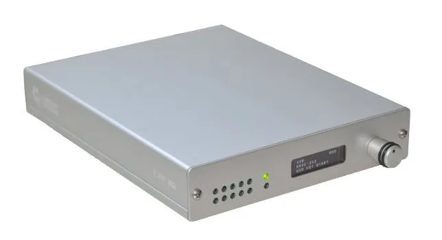

To register motor responses, we use the system «932 fORP» (Current Designs, USA), which includes two four-button consoles and a trackball.
The opto-electronic interface 932 receives optical signals from handheld devices in the MR environment and converts them into electronic signals for the computer.
We also use various fiber-optic devices developed by the specialists of the Center for specific experiments.
Movement Registration
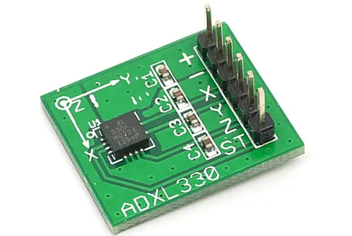
To record subject’s movements, we use three-axis accelerometers «iMEMS ADXL330» (Analog Devices, USA). The device can measure accelerations up to ±4 g along three spatial axes in analog form.
Triggers
To synchronise stimulation events and subject’s responses with the MEG data, we use two 16-channel analog-digital receivers.
Biological Feedback
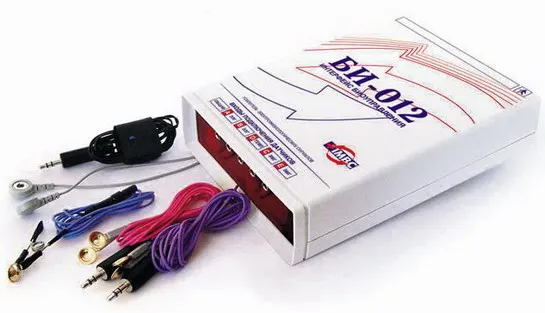
The «Бослаб БИ-012» hardware-software communication system enables biofeedback sessions, psychophysiological diagnostics, and various kinds of trainings based on biological feedback. The biological feedback principle is widely used to correct a range of central nervous system dysfunctions.
Software

Proprietary software
«MaxFilter 2.2» (Electa Neuromag, Finland, User’s Manual) performs:
- Removal of noise originating from sources outside the sensor array;
- Detection of bad channels;
- Compensation for head movements (with continuous HPI-monitoring);
- Data transformation to standard space (e.g., for intersubject data alignment).
«Presentation» (Neurobehavioral Systems, USA):
- To control experiments: presents auditory, visual, and multimodal stimuli with sub-millisecond timing precision.
Open-source software
FreeSurfer for MRI data processing and analysis:
- Automatic segmentation and reconstruction of cerebral cortex;
- Generation of 3D models of brain anatomy;
- Cortical thickness analysis and morphometric studies.
MNE-Python, Brainstorm for MEG and EEG data processing and analysis.
PsychoPy for creating behavioral experiments.
The MEG-center is also equipped with technical tools for preparation of experiments, as well as with diagnostic equipment (e.g. «WaveAce 222» oscilloscope (LeCroy, USA, 2GS/s), signal generator «АКИП-3402» (Prist, Russia), a soldering station etc.).